What Is a PVDF Membrane and How Is It Used in the Laboratory?
By LydiaPosted on June 30, 2025Category: Hollow Fiber
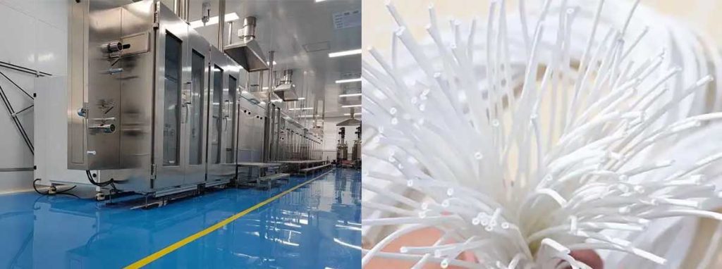
A PVDF membraneconsists of a synthetic polymer with a hydrophobic, microporous structure, making it a preferred choice in the laboratory for filtration and protein detection. This membrane offers high protein binding capacity, robust chemical resistance, and strong mechanical strength. Researchers often pre-activate the membrane by soaking it in methanol, which greatly improves buffer infiltration and protein transfer. The hollow fiber spinning machine frequently produces this membrane, ensuring uniform pore distribution. In laboratory detection tasks, the membrane demonstrates reliable performance, especially with high molecular weight proteins, as shown by comparative studies of binding capacity.
Key Takeaways
PVDF membranes have strong hydrophobic surfaces that bind proteins well, making them ideal for protein detection methods like Western blotting.
These membranes resist many chemicals and high temperatures, allowing reliable use in harsh laboratory conditions and filtration tasks.
PVDF membranes offer excellent mechanical strength, so they stay intact during handling and repeated use in experiments.
PVDF syringe filters help remove particles and bacteria from samples while preserving sample quality due to low protein binding and chemical resistance.
Proper storage and activation, such as soaking in methanol before use, improve membrane performance and ensure consistent, reliable laboratory results.
PVDF Membrane Properties
Hydrophobicity
A key feature of the pvdf membrane is its strong hydrophobic nature. This property means that the membrane repels water, which makes it highly effective for filtering non-polar substances and for applications where water resistance is essential. The hydrophobic polyvinylidene fluoride membrane stands out in laboratory settings because it can prevent unwanted water absorption, maintaining consistent performance during filtration and protein transfer.
Researchers often measure hydrophobicity by examining how much protein or other molecules adhere to the membrane surface. The following table compares the adsorption of bovine serum albumin (BSA) and other molecules on different membrane types:
Measurement Type
Membrane Type
Quantitative Result
Interpretation
BSA adsorption (QCM-D method)
PVDF
Highest amount adsorbed
Indicates more hydrophobic surface prone to fouling
BSA adsorption (QCM-D method)
EVOH (hydrophilic)
Lowest amount adsorbed
Indicates hydrophilic surface resists fouling
BSA adsorption (SPR method)
PVDF/PMMA-g-PEO9
Adsorbed ~50% of BSA compared to pure PVDF
Shows enhanced fouling resistance due to hydrophilicity
Dextran adsorption (SPR method)
PVDF/PMMA-g-PEO9
Adsorbed ~9% (one-eleventh) compared to pure PVDF
Demonstrates significant reduction in fouling
Contact angle measurement
PVDF/DA modified membrane
Contact angle ~33.9° (lower than pure PVDF)
Indicates increased hydrophilicity and reduced hydrophobicity
Pure water flux (PVDF/DA)
PVDF/DA membrane
126.24 mL/(cm²·h) at 0.10 MPa
Reflects improved membrane performance linked to surface properties
This data shows that the pvdf membrane’s hydrophobicity can lead to higher protein adsorption, which is useful for certain detection methods but may also increase the risk of fouling. Modifications to the membrane surface can adjust this property for specific laboratory needs.
Protein Binding
The pvdf membrane is well known for its high protein binding capacity. This characteristic is especially important in laboratory detection techniques such as Western blotting, where the membrane must capture and retain proteins for visualization. Studies show that pvdf membrane can bind between 170 and 200 μg of protein per square centimeter. In comparison, nitrocellulose membranes bind only 80 to 100 μg/cm². This higher binding capacity allows for greater sensitivity in protein detection, making the pvdf membrane a preferred choice for researchers who need to detect low-abundance proteins. However, the strong binding can sometimes result in higher background signals, so careful optimization is necessary during antibody-based detection.
Chemical and Thermal Resistance
The pvdf membrane demonstrates excellent resistance to a wide range of chemicals and high temperatures. This property allows the membrane to perform reliably in harsh laboratory environments, including those involving organic solvents or elevated temperatures. For example, when researchers incorporate silver nanoparticles into the membrane and apply thermal treatment, the membrane resists degradation up to 465 °C, compared to 370 °C for standard pvdf membranes. No silver leaching occurs during filtration, confirming the chemical stability of the membrane. Blending pvdf with fluorinated polyimide further improves resistance to organic solvents and maintains performance at temperatures up to 90 °C. The high bond energy of carbon-fluorine bonds in the membrane structure contributes to this durability. These features make the pvdf membrane suitable for demanding applications such as wastewater treatment, high-temperature filtration, and laboratory detection protocols that require exposure to aggressive chemicals.
Mechanical Strength
Mechanical strength is another advantage of the pvdf membrane. The membrane must withstand physical stress during handling, filtration, and detection procedures. Tensile tests show that composite pvdf membranes can reach ultimate tensile strengths up to 31.55 MPa, with increased Young’s modulus values when reinforced with filler particles like barium titanate or bacterial nanocellulose. The following table summarizes the mechanical properties of different pvdf membrane compositions:
PVDF Membrane Composition
Average Yield Strength (MPa)
Average Elongation at Break (%)
15 wt% PVDF + 1% SnO2
0.488
2.03
17.5 wt% PVDF + 1% SnO2
0.649
2.11
20 wt% PVDF + 1% SnO2
0.746
3.75
Membranes produced using the hollow fiber spinning machine often display enhanced mechanical properties due to their uniform structure. Some pvdf membranes prepared by vapor-induced phase separation achieve tensile stress at break values above 4.0 MPa and tensile strain around 33%. This strength ensures that the membrane remains intact during repeated use and under various laboratory conditions.
Tip: The combination of hydrophobicity, high protein binding, chemical and thermal resistance, and mechanical strength makes the pvdf membrane a versatile tool in laboratory detection and filtration. Researchers can select or modify the membrane to match the specific requirements of their experiments.
Laboratory Uses
Western Blotting
Western blotting remains one of the most common applications for the pvdf membrane in the laboratory. Scientists use western blot membranes to transfer proteins from gels onto a solid support, allowing for precise protein detection. The pvdf membrane offers high protein binding capacity, which is essential for capturing proteins during the transfer step. Researchers often pre-activate the membrane with methanol to improve protein transfer efficiency. This step ensures that the membrane becomes more receptive to buffer solutions and proteins, which is critical for a successful western blotting protocol.
Statistical comparisons show that pvdf membrane outperforms nitrocellulose membranes when detecting high molecular weight proteins. In slot blot assays, pvdf membrane provides reliable results with strong signal intensity and low background noise. The following table summarizes key findings from comparative studies:
Lectins and antibodies in multiple re-probing scenarios
n=3; statistical significance with p-values <0.05 and <0.01; error bars
PVDF membrane shows superior re-probing capacity compared to NC membrane in all tested cases.
Direct protein binding (slot blot style)
PVDF vs NC
Trypsin, BSA, Fetuin, Lactase
n=3; statistical analysis; error bars
NC membrane binds low molecular weight proteins better; PVDF shows saturation or decline at high concentrations.
Researchers value the pvdf membrane for its ability to support multiple rounds of probing and stripping, which is important for complex protein analysis workflows. Western blot membranes made from pvdf also maintain their integrity during repeated handling, thanks to their mechanical strength.
PVDF Syringe Filters
PVDF syringe filters play a vital role in laboratory filtration. Scientists use these filters to remove particulates, bacteria, and other contaminants from liquid samples. The pvdf membrane inside these filters provides excellent chemical resistance, allowing filtration of aggressive solvents, acids, and bases without degradation. This property makes pvdf syringe filters suitable for a wide range of laboratory applications, including sterile filtration and hplc sample preparation.
Key features of pvdf syringe filters include:
Low protein binding, which minimizes analyte loss and preserves sample integrity.
Consistent pore size, ensuring high particle retention efficiency.
Compatibility with sterilization methods such as autoclaving and gamma irradiation.
Hydrophobic nature, which prevents water absorption and reduces analyte loss during hplc sample preparation.
Effective removal of bacteria and particulates with common pore sizes (0.2 μm and 0.45 μm).
Scientists rely on several methods to verify the performance of pvdf syringe filters:
Particle counting before and after filtration to measure efficiency.
Microbial filtration tests to determine removal rates of microorganisms.
HPLC analysis to check for retention or loss of target molecules.
Control of variables such as sample volume, filtration speed, and membrane pore size for accurate results.
Tip: Proper use of pvdf syringe filters helps maintain sample purity and protects analytical instruments from damage caused by particulates.
PVDF syringe filters also support environmental sample filtration, enabling accurate contaminant analysis in complex matrices. Their robust design ensures consistent performance, even under high pressure or flow rates.
Sample Preparation
Sample preparation is a critical step in many laboratory workflows, especially for protein analysis and filtration. The pvdf membrane finds extensive use in this area due to its controlled pore size and strong mechanical properties. Scientists use pvdf filter membranes in immunoassays, lateral flow tests, and hplc sample preparation. The membrane’s structure, often produced by the hollow fiber spinning machine, ensures uniformity and reliability.
Experimental studies show that pvdf membrane created by vapor- and non-solvent-induced phase separation offers suitable capillary flow rates and mechanical strength. These properties are essential for applications like colloidal gold test strips and immunoassays. Although the protein adsorption capacity of these membranes is lower than that of commercial nitrocellulose membranes, they still perform effectively in sample preparation for analytical testing methods.
Researchers also use pvdf syringe filters during hplc sample preparation to remove particulates and ensure sample clarity. The hydrophobic nature of the membrane prevents water absorption, which helps maintain consistent analytical results.
Gravimetric and Microanalysis
Gravimetric and microanalysis require precise and reliable filtration. The pvdf membrane supports these applications by providing high chemical resistance and mechanical durability. Scientists use the membrane to collect and weigh particulates from liquid samples, ensuring accurate measurements. In membrane-assisted crystallization experiments, composite pvdf membranes with 2D nanofillers such as graphene and bismuth telluride outperform pristine pvdf membranes.
The following table highlights the performance of different pvdf membranes in NaCl crystallization:
Membrane Type
Time to First Visible Crystals (min)
Membrane Flux (L·m⁻²·h⁻¹)
Contact Angle (°)
Thickness (μm)
Mean Pore Size (μm)
Porosity (%)
Crystal Size (μm)
Crystal Count (B0)
Crystal Size Uniformity (CV %)
Pristine PVDF
285
N/A
139 ± 3
71 ± 2
0.52 ± 0.05
82 ± 4
20.6
490,593
77.1
PVDF/BT (0.5%)
140
2.7
128 ± 8
68 ± 1
0.50 ± 0.2
75 ± 1
9.56
1,060,979
N/A
PVDF/BT (7%)
N/A
3.9
130 ± 2
100 ± 5
0.50 ± 0.08
77 ± 1
13.38
337,756
47
PVDF/G (0.5%)
N/A
1.6
136 ± 1
62 ± 3
0.24 ± 0.05
56 ± 7
18.92
280,475
36.7
Composite pvdf membranes reduce nucleation time and promote more uniform and numerous NaCl crystal formation. These improvements support more accurate gravimetric analysis and microanalysis in laboratory settings.
Note: The combination of high chemical resistance, mechanical strength, and controlled pore size makes the pvdf membrane a trusted choice for gravimetric and microanalysis, sterile filtration, and other demanding laboratory filtration tasks.
Handling and Best Practices
Compatibility
Scientists select the right membrane based on compatibility with solvents and buffers. The pvdf membrane resists many chemicals, including acids, bases, and organic solvents. This resistance makes it ideal for laboratory filtration and detection tasks. However, some strong oxidizers or concentrated acids can damage the membrane. Researchers often use pvdf syringe filters for samples containing aggressive chemicals. These filters maintain integrity during filtration, even when exposed to harsh solvents. The hydrophilic polyvinylidene fluoride membrane offers better performance with aqueous solutions. The hollow fiber spinning machine produces membranes with consistent pore size, which helps ensure compatibility across different applications.
Tip: Always check the chemical compatibility chart before using a membrane with unfamiliar solvents.
Storage
Proper storage extends the life of the pvdf membrane and pvdf syringe filters. Scientists store unused membranes in a cool, dry place away from direct sunlight. They keep membranes in their original packaging until use to prevent contamination. For pvdf syringe filters, sealed packaging protects the membrane from dust and moisture. After opening, users should reseal the package or use a clean container. Membranes exposed to air for long periods may lose performance. Researchers avoid storing membranes near strong acids or bases to prevent chemical damage.
Storage Checklist:
Keep membranes dry and cool
Avoid sunlight and heat
Store in original or airtight packaging
Separate from reactive chemicals
Activation Steps
Before use in protein detection, scientists activate the pvdf membrane by soaking it in methanol. This step changes the membrane from hydrophobic to hydrophilic, allowing better buffer penetration. For pvdf syringe filters, users may pre-wet the membrane with methanol or water, depending on the application. Activation ensures efficient protein transfer and reduces background noise in detection. Skipping this step can lead to poor results or incomplete filtration.
Note: Always follow the manufacturer’s instructions for activation. Methanol is flammable, so handle it with care.
Researchers maximize membrane performance by following these best practices. They choose the right membrane for each task, store it properly, and activate it before use. These steps help maintain the quality of pvdf syringe filters and ensure reliable results in laboratory detection.
Conclusion
PVDF membrane offers laboratories a unique combination of chemical resistance, thermal stability, and mechanical strength. Researchers value the membrane for its biocompatibility, broad pH range, and efficient filtration. The membrane’s versatility extends from protein detection to wastewater treatment, with the hollow fiber spinning machine ensuring consistent membrane quality. While the membrane may have higher costs and some limitations compared to PTFE, its low protein binding and sterilizability make it ideal for sensitive applications. Understanding membrane properties helps scientists select the right membrane for each task. For advanced membrane troubleshooting or application tips, consult specialized resources.


